Information
Chemical Name: (4S)-2-(6-hydroxy-1,3-benzothiazol-2-yl)-4,5-dihydrothiazole-4-carboxylic
CAS No.: 115144-35-9
Molecular Structure: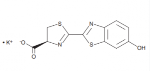
Molecular Formula: C11H8N2O3S2K
Molecular Weight:318.41
Purity: ≥98%
Appearance: Yellow powder
Storage Condition: Store at -20℃, protect from light
Shipment:
Description
D-Luciferin is a common substrate for Luciferase used throughout the biotechnologyand specifically for in vivo imaging. The water soluble substrate for the firefly luciferase enzyme utilizes ATP and Mg2+ as cofactors to emit a characteristic yellow-green emission in the presence of oxygen.Through the utilization of ATP, the reaction can be further used to indicate the presence of energy or life in order to function as a life-death stain.

Luciferase labeled tumor cells, stem cells or infectious diseases are often inoculated into research animals such as rats or mice for investigation. The injection of luciferin allows for the real-time, noninvasive monitoring of disease progression and/or drug efficacy in these model systems through Bioluminescence Imaging (BLI).
Luciferin is also commonly used for in vitro research, including luciferase and ATP assays, gene reporter assays, high throughput sequencing and various contamination assays.
Performance
1. Structural determination (HNMR)
H-NMR spectrum, from the above spectrum, it can be seen that the product structure of MeilunBio is correct.
2. Purity testing (HPLC)
HPLC spectrum, from the above spectrum, it can be seen that the purity of the product is very high.
3. Verification of fluorescence intensity

Figure A: D-luciferin K signal intensity detection
The BioTek Synergy HTX was used to verify meilunstar D-luciferin K and company P’s. Double-blind control results showed that there was no significant difference in fluorescence intensity between the two.

Figure B: D-Luciferin K fluorescence signal stability (kinetics) detection.
Equal amounts of firefly luciferase were added to the classic firefly luciferase luminescence buffer solution (containing ATP, CoA, Mg2+) with 300μg/ml D-Luciferin K, and the fluorescence signal was continuously tracked for 60s from the initial moment of the reaction. It can be found that signal stability of meilunstar D-Luciferin K is not significantly different from those of imported companies. (Instrument: BioTek HTX)
4. Solubility Test
At room temperature, use pure water as the solvent to test the solubility. The concentration is 30MG/ML. It is easy to dissolve and has good clarity.

5. In vivo imaging examples

6. In vitro bioluminescence experiments
Prepare materials:
D-luciferin potassium salt (Cat No., MB1834)
Sterile water (Cat No., MA0028)
Complete culture medium (self-configured)
Steps:
- Adherent cells: Inoculate the cells in culture plates and incubate for several hours or overnight to make the cells adherent to the wall. Suspended cells: the cells can be inoculated into the culture plate and then directly followed up without incubation. If the doubling time is relatively short, the doubling time should be considered for cell counting.
- Prepare 100X luciferin stock solution (15mg/ml) by dissolving 15mg of Fluorescein Potassium Salt in 1ml of sterile water, gently invert and shake until Fluorescein Potassium Salt is completely dissolved. Use immediately after mixing or freeze at -20℃ after dispensing.
- Dilute 100X luciferin stock solution (15mg/ml) with pre-warmed cell culture medium 1:100 to formulate luciferin working solution (final concentration 150μg/ml), ready to use.
- Remove the culture medium from the culture plate.
- Add appropriate amount of luciferin working solution into the cells before imaging, and then perform image analysis. Note: Incubate the cells at 37℃ for a short time before imaging to enhance the signal. The incubation time depends on the specific cell type. Generally 10 minutes of incubation is sufficient, test and adjust as needed.
- Every 10 minutes, up to a maximum of 40 minutes, examine in vitro bioluminescence using the VILBER FUSION FX Imaging System to determine the kinetic profile and identify peak time points for the cells.

Figure: D-luciferin K in vitro bioluminescence kinetics curve
Use MeilunStar D-luciferin K to detect the 293T cells transfected with pML-NFκB-Fluc2-Neo plasmid (MailunBio Catalog No. MA0504), detect the fluorescent signal every 5 minutes, and continue to track for 40 minutes. The kinetic curve shows that the bioluminescence of cells using MeilunStar D-luciferin K as a substrate decays slowly, and the fluorescence signal intensity only decays by about 25% after 40 minutes.
Usage Statement
Research Use Only (RUO)
All sales are subject to the General Terms and Conditions of Sale set forth on our website.