Product Description
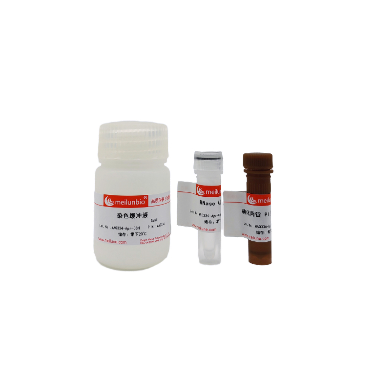
The Cell Cycle and Apoptosis Analysis Kit used classic PI staining methods to detect cell cycle and apoptosis through flow cytometry.
The cell cycle refers to the whole process that a cell goes through from the completion of one division to the end of the next, during which the cell’s genetic material replicates and doubles, and is evenly divided into two daughter cells at the end of division. The cell cycle is divided into two stages: interphase and division phase (M phase), and the interphase is further divided into: dormant phase (G0), early DNA synthesis phase (G1 phase), DNA synthesis phase (S phase) and late DNA synthesis phase (G2 phase). Using the characteristics of the DNA in the cell that can bind to fluorescent dyes (such as Propidium Iodide), the DNA content of the cell at each stage is different, and the fluorescence intensity detected is different, so as to reflect the status of the cell cycle at each stage, that is, the cell proliferation status.
Propidium Iodide (PI) is a fluorescent dye that can be inserted into and bound to the base pairs of double-stranded DNA and RNA without base specificity. PI can produce fluorescence after binding with double-stranded DNA, and the fluorescence intensity is proportional to the content of double-stranded DNA. After the DNA in the cells is stained by PI, the DNA content of the cells can be determined by flow cytometry, so as to analyze the cell cycle and apoptosis. Assuming that the fluorescence intensity of G0/G1 phase cells is 1, the theoretical value of the fluorescence intensity of G2/M phase cells containing double genomic DNA is 2, and the fluorescence intensity of S phase cells undergoing DNA replication is between 1-2.
Due to nucleus concentration and DNA fragmentation, some genomic DNA fragments of apoptotic cells were lost during the staining process. Therefore, apoptotic cells showed obvious weak staining after Propidium Iodide staining, that is, the fluorescence intensity was less than 1, and the so-called sub-G1 peak appeared in the fluorescence pattern detected by flow detection. The apoptotic cell peak. PI staining can also distinguish between cell peak types of apoptosis and cell necrosis. During apoptosis, apoptotic cells are wrinkled, chromatin is concentrated, nuclear fragmentation is produced, apoptotic bodies are produced, and the forward angular light scattering (FSC) of cells is lower than normal. In the early stages of apoptosis, the ability of cells to scatter forward angular light (FSC) is significantly reduced, and the ability to scatter lateral angular light (SSC) is increased or unchanged. Both FSC and SSC signaling decreased in the late stage of apoptosis. When the cells die, the cells mostly show swelling, so the FSC is higher than normal, and the SSC is higher than normal.
This kit is suitable for cell cycle and apoptosis detection of cultured adherent or suspended cells, and can also be used to distinguish apoptosis from cell necrosis. When used for cell cycle and apoptosis detection of tissue, the tissue must be digested into a single-cell state. The kit detects the cell content of each sample in the range of 105-106, which is enough to test 50 samples.
Performance
Cell cycle assays
Apoptosis-inducing cells versus normal cell cycle fitting plot. Good cycle curve fit and reasonable data.
Shipping and Storage
- Storage:Stored at -20°C, valid for one year. Propidium Iodide PI Staining Solution (20x) needs to be stored away from light. The kit can be stored at 4°C, valid for one month
- Shipment:
Usage Statement
Research Use Only (RUO)
All sales are subject to the General Terms and Conditions of Sale set forth on our website.


