Product Description
The EdU Cell Proliferation Kit with Alexa Fluor 488 is a reagent kit that utilizes nucleoside incorporation for rapid, simple, and highly sensitive detection of cell proliferation. The principle is as follows: EdU (5-ethynyl-2′-deoxyuridine) included in the kit is a thymidine analog. During the S phase of the cell cycle, EdU substitutes for thymidine in the newly synthesized DNA. Additionally, the ethynyl group on EdU can undergo a covalent reaction with azides (such as the fluorescent probe Alexa Fluor 488 Azide) catalyzed by copper ions (Cu+), forming a stable triazole ring in a reaction known as the click reaction (See Figure 1). This reaction rapidly labels the newly synthesized DNA with green fluorescence, allowing for the detection of cell proliferation by measuring the fluorescence signal.
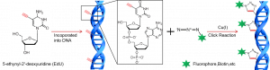
Figure 1. Schematic diagram of the click reaction in the EdU method.
Cell proliferation assays are fundamental experimental methods for assessing cell viability, genetic toxicity, and the efficacy of anti-cancer drugs. Cell proliferation detection methods are typically categorized into five types: membrane damage detection, metabolic activity measurement, ATP level determination, DNA synthesis detection, and cell fluorescence labeling methods. The most precise method recognized currently for detecting cell proliferation is the direct detection of DNA synthesis in cells, namely the nucleoside incorporation method. The previously common nucleoside incorporation method was the BrdU (bromodeoxyuridine, a thymidine nucleotide analog) method. However, a drawback of the BrdU method is that it requires DNA denaturation before it can bind to antibodies, which disrupts the DNA double-helix structure and affects the binding of other dyes, leading to dispersed staining and reduced accuracy. The EdU method has the following advantages:
- Safety: Non-radioactive, avoids the use of [3H]thymidine.
- Simplicity: Based on a simple chemical reaction, requires only three steps: EdU incubation, cell fixation, and fluorescence detection. Does not require DNA denaturation or antibody incubation.
- Speed: The entire detection process takes only 2.5 hours, significantly reducing the experimental timeframe.
- Accuracy: High labeling efficiency without the need for DNA denaturation, minimizing sample damage and ensuring clear nuclear margins.
- Sensitivity: Does not require antibodies; the detection dye is 1/500 the concentration of BrdU antibodies, facilitating easier diffusion and accurate detection of even single proliferating cells.
- Compatibility: Minimal sample damage allows simultaneous labeling with various antibodies or fluorescent proteins.
This kit is suitable for cultured cells or tissue samples, as well as for tissue sections. It allows for the detection of individual proliferating cells within cell or tissue samples and facilitates quantitative analysis of overall cell proliferation. The kit includes all components required for the EdU detection method and provides the blue nuclear stain Hoechst 33342, which can be used to restain all nuclei and is also useful for cell cycle analysis. Results obtained with this kit can be examined using a fluorescence microscope, laser confocal microscope, flow cytometer, or fluorescence plate reader, and are applicable to high-content screening.
For cells cultured in 6-well plates (500 μl of Click reaction solution per well), the kit provides enough reagents for 50 wells; for cells in 96-well plates (50 μl of Click reaction solution per well), it can supply reagents for 500 wells. The specific volume of Click reaction solution for different container types can be found in Table 1. For flow cytometry with 100,000 to 1,000,000 cells per tube (500 μl of Click reaction solution per tube), the kit can support reactions in 50 tubes; for frozen or paraffin-embedded sections (100-200 μl of Click reaction solution per sample), it can provide enough for 125-250 sample reactions.
Spectral characteristics: 488-Azide: green fluorescence, Ex/Em=491/518 nm; Hoechst 33342: blue fluorescence, Ex/Em=346/460 nm, bound to DNA.
Performance
1. Flow cytometry of suspension cells
Jurkat cells were labeled with EdU only (left column) or treated with 10 mM DNA synthesis inhibitor Hydroxyurea for 30 min prior to labeling with EdU (right column), stained 2 h later, and then detected by flow cytometry.
As can be seen from the graph, only EdU-labeled cells had a higher proportion of green fluorescence-positive cells, showing two peaks of green fluorescence negative (weak staining) and positive (strong staining), corresponding to unproliferating and proliferating cells, respectively. In Hydroxyurea-treated cells, the green fluorescence-positive cells almost completely disappeared, leaving only one green fluorescence-negative peak.
2. Fluorescence microscopy of adherent cells
Hela cells were labeled with EdU only (left column), or treated with 10 mM DNA synthesis inhibitor Hydroxyurea for 30 min before labeling with EdU (right column), stained 2 h later (the step was stained with Hoechst at the end, which was convenient for observing the proportion of proliferating cells), and then examined by fluorescence microscopy.
Under the microscope, only EdU-labeled cells had a portion of proliferating cells stained with green fluorescence, the nuclei of non-proliferating cells were single-stained with blue color, and the nuclei of proliferating cells were double-stained with green and blue color; in the Hydroxyurea-treated cells, there were almost no green-fluorescent proliferating cells, and the nuclei of the vast majority of the cells were only blue in color.
Shipping and Storage
- Storage:Store at -20°C, effective for 12 months.488-Azide and Hoechst 33342 (1000X) must be protected from light.
- Shipment:
Usage Statement
Research Use Only (RUO)
All sales are subject to the General Terms and Conditions of Sale set forth on our website.